Thành viên:Naazulene/Bộ xương người
| Human skeleton | |
|---|---|
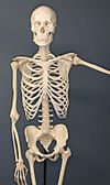
 A human skeleton on exhibit at the Museum of Osteology, Oklahoma City, Oklahoma | |
| Chi tiết | |
| Định danh | |
| Tiếng Hy Lạp | σκελετός |
| Thuật ngữ giải phẫu | |
Bộ xương người là cấu trúc khung của cơ thể người. Em bé mới sinh có khoảng 270 xương, nhưng các xương dung hợp với nhau khi con người phát triển và người trưởng thành chỉ có 206 xương.[1] Khối lượng của bộ xương (khoảng 10 - 11kg) chiếm 14% tổng khối lượng cơ thể và đạt khối lượng tối đa trong độ tuổi 25 đến 30.[2] Bộ xương người có thể được chia thành xương trục và xương treo / xương chi. Xương trục bao gồm cột sống, lồng ngực, sọ và một số xương khác. Xương treo là nhưng xương gắn vào xương trục, bao gồm đai chậu, đai vai và xương tay, xương chân.
Bộ xương người thực hiện sáu chức năng chính: nâng đỡ, cử động, bảo vệ, trữ khoáng, tạo huyết, và điều hòa nội tiết.
Bộ xương người không khác biệt ở nam và nữ như một số linh trưởng khác, nhưng vẫn có một số khác biệt nhỏ ở sọ, răng, xương dài và xương chậu. Nhìn chung, các yếu tố xương ở người nữ thường nhỏ và ít cường tráng hơn so với người nam trong cùng quần thể; xương chậu của người nữ khác người nam để thuận tiện sinh đẻ.[3] Khác với hầu hết các loài sinh trưởng, dương vật người không có xương.[4]
Phân loại
sửaXương trục
sửaBộ xương trục (80 xương) bao gồm cột sống (32 - 34 xương; số lượng khác nhau giữa người với người và khác nhau ở xương cùng và xương cụt), một phần khung sườn (12 cặp xương sườn và xương đòn), và xương sọ (22 xương và 7 xương liên quan)
Tư thế đứng thẳng được nâng đỡ bởi xương trục bằng cách chuyển trọng lượng từ đầu và thân mình xuống hông và chân. Những xương của cột sống được hỗ trợ bởi nhiều dây chằng. Cơ dựng cột sống cũng giúp nâng đỡ và thăng bằng.
Xương treo
sửaBộ xương treo (126 xương) bao gồm đai ngực, chi trên, đai chậu, và chi dưới. Chức năng của chúng là vận động và bảo vệ các cơ quan tiêu hóa, bài tiết, và sinh sản.
Chức năng
sửaBộ xương có sáu chức năng chính: nâng đỡ, cử động, bảo vệ, tạo huyết, trữ khoáng, và điều hòa nội tiết.
Nâng đỡ
sửaBộ xương là cấu trúc khung để nâng đỡ cơ thể và giữ hình dáng của nó. Xương chậu, cùng với những cơ và dây chằng gắn vào, tạo nên sàn chậu là nền của những cấu trúc chậu. Mặt khác, nếu không có khung sườn, sụn sườn, và cơ gian sườn, phổi sẽ sụp.
Cử động
sửaKhớp gắn các xương lại với nhau và cho phép sự cử động, một số khớp cho phép nhiều cử động hơn khớp khác, ví dụ khớp ổ - cầu cho phép nhiều cử động hơn là khớp xoay ở cổ. Sự cử động được thực hiện bởi cơ xương, là những cơ gắn vào các vị trí khác nhau ở xương. Cơ, xương, và khớp hình thành nên cơ chế chung cho cử động, dưới sự điều khiển của hệ thần kinh.
Người ta cho rằng mật độ xương giảm thì sự nhanh nhẹn và khéo léo của cử động cũng giảm. Người tiền sử có mật độ xương cao hơn hẳn vì lối sống săn bắt bị thay thế bởi nông nghiệp.[5][6][7]
Bảo vệ
sửaXương bảo vệ nhiều nội quan quan trọng khỏi chấn thương:
- Hộp sọ bảo vệ não
- Cột sống bảo vệ tủy sống
- Khung sườn, cột sống, xương đòn bảo vệ phổi, tim, và các mạch máu lớn
Tạo huyết
sửaXương, cụ thể là tủy xương là địa điểm tạo huyết - sản xuất tế bào máu. Ở trẻ em, sự tạo huyết diễn ra chủ yếu ở tủy xương của xương dài như xương đùi và xương cánh tay. Ở người lớn, nó diễn ra chủ yếu ở xương chậu, xương sọ, xương sống và xương ức.[8]
Trữ khoáng
sửaChất cơ bản (bone matrix) của xương có thể trữ calci nên nó tham gia vào quá trình chuyển hóa calci. Tủy xương có thể trữ sắt trong ferritin nên nó tham gia vào quá trình chuyển hóa sắt. Tuy nhiên, xương không phải chỉ có calci, mà là một hỗn hợp chondroitin sulfate và hydroxyapatite, trong đó hydroxyapatite chiếm 70% xương. Hydroxyapatite chứa 39,8% calci, 41,4% oxygen, 18.5% phospho và 0.2% hydro (tỉ lệ khối lượng). Chondroitin sulfate là một loại đường được cấu tạo chủ yếu bởi oxygen và carbon.
Điều hòa nội tiết
sửaTế bào xương tiết ra một hormone tên là osteocalcin có khả năng điều hòa đường huyết và sự lắng đọng chất béo. Osteocalcin làm tăng tiết insulin và sự nhạy cảm đối với insulin, cùng với tăng số lượng tế bào tiết insulin và làm giảm sự trữ chất béo.[9]
Khác biệt giới tính
sửaKhác biệt giải phẫu giữa người nam và người nữ rất rõ ràng ở một số phần mềm, nhưng không quá nhiều ở xương. Tuy nhiên, vẫn có một số khác biệt nhỏ ở sọ, răng, xương dài và xương chậu. Nhìn chung, những yếu tố xương ở người nữ thường nhỏ và ít cường tráng hơn ở người nam của cùng quần thể. Người ta vẫn chưa thể khẳng định những khác biệt đó là do gen hay do môi trường.
Xương sọ
sửaNhững cấu trúc mang sự khác biệt là đường gáy giữa, mõm chũm, cung hốc mắt, và cằm.[10]
Răng
sửaKhác biệt chủ yếu ở răng nanh, nhưng cũng không khác dữ như ở những loài vượn lớn khác.
Xương dài
sửaXương dài ở người nam thường dài hơn ở người nữ của cùng quần thể. Cơ bám vào cũng chặt hơn ở người nam.
Long bones are generally larger in males than in females within a given population. Muscle attachment sites on long bones are often more robust in males than in females, reflecting a difference in overall muscle mass and development between sexes. Sexual dimorphism in the long bones is commonly characterized by morphometric or gross morphological analyses.
Pelvis
sửaThe human pelvis exhibits greater sexual dimorphism than other bones, specifically in the size and shape of the pelvic cavity, ilia, greater sciatic notches, and the sub-pubic angle. The Phenice method is commonly used to determine the sex of an unidentified human skeleton by anthropologists with 96% to 100% accuracy in some populations.[11]
Women's pelvises are wider in the pelvic inlet and are wider throughout the pelvis to allow for child birth. The sacrum in the women's pelvis is curved inwards to allow the child to have a "funnel" to assist in the child's pathway from the uterus to the birth canal.
Ý nghĩa lâm sàng
sửaCó rất nhiều rối loạn về xương, phổ biến nhất là loãng xương và vẹo cột sống.
Viêm khớp
sửaArthritis is a disorder of the joints. It involves inflammation of one or more joints. When affected by arthritis, the joint or joints affected may be painful to move, may move in unusual directions or may be immobile completely. The symptoms of arthritis will vary differently between types of arthritis. The most common form of arthritis, osteoarthritis, can affect both the larger and smaller joints of the human skeleton. The cartilage in the affected joints will degrade, soften and wear away. This decreases the mobility of the joints and decreases the space between bones where cartilage should be.
Loãng xương
sửaOsteoporosis is a disease of bone where there is reduced bone mineral density, increasing the likelihood of fractures.[12] Osteoporosis is defined by the World Health Organization in women as a bone mineral density 2.5 standard deviations below peak bone mass, relative to the age and sex-matched average, as measured by dual energy X-ray absorptiometry, with the term "established osteoporosis" including the presence of a fragility fracture.[13] Osteoporosis is most common in women after menopause, when it is called "postmenopausal osteoporosis", but may develop in men and premenopausal women in the presence of particular hormonal disorders and other chronic diseases or as a result of smoking and medications, specifically glucocorticoids.[12] Osteoporosis usually has no symptoms until a fracture occurs.[12] For this reason, DEXA scans are often done in people with one or more risk factors, who have developed osteoporosis and be at risk of fracture.[12]
Osteoporosis treatment includes advice to stop smoking, decrease alcohol consumption, exercise regularly, and have a healthy diet. Calcium supplements may also be advised, as may vitamin D. When medication is used, it may include bisphosphonates, strontium ranelate, and osteoporosis may be one factor considered when commencing hormone replacement therapy.[12]
History
sửaIndia
sửaSuśruta-saṃhitā, composed between 6th century BCE and 5th century CE speaks of 360 bones. Books on Salya-Shastra (surgical science) know of only 300. The text then lists the total of 300 as follows: 120 in the extremities (e.g. hands, legs), 117 in the pelvic area, sides, back, abdomen and breast, and 63 in the neck and upwards.[14] The text then explains how these subtotals were empirically verified.[15] The discussion shows that the Indian tradition nurtured diversity of thought, with Sushruta school reaching its own conclusions and differing from the Atreya-Caraka tradition.[15] The differences in the count of bones in the two schools is partly because Charaka Samhita includes thirty two teeth sockets in its count, and their difference of opinions on how and when to count a cartilage as bone (both count cartilages as bones, unlike current medical practice).[16]
Hellenistic world
sửaThe study of bones in ancient Greece started under Ptolemaic kings due to their link to Egypt. Herophilos, through his work by studying dissected human corpses in Alexandria, is credited to be the pioneer of the field. His works are lost but are often cited by notable persons in the field such as Galen and Rufus of Ephesus. Galen himself did little dissection though and relied on the work of others like Marinus of Alexandria,[17] as well as his own observations of gladiator cadavers and animals.[18] According to Katherine Park, in medieval Europe dissection continued to be practiced, contrary to the popular understanding that such practices were taboo and thus completely banned.[19] The practice of holy autopsy, such as in the case of Clare of Montefalco further supports the claim.[20] Alexandria continued as a center of anatomy under Islamic rule, with Ibn Zuhr a notable figure. Chinese understandings are divergent, as the closest corresponding concept in the medicinal system seems to be the meridians, although given that Hua Tuo regularly performed surgery, there may be some distance between medical theory and actual understanding.
Renaissance
sửaLeonardo da Vinci made studies of the skeleton, albeit unpublished in his time.[21] Many artists, Antonio del Pollaiuolo being the first, performed dissections for better understanding of the body, although they concentrated mostly on the muscles.[22] Vesalius, regarded as the founder of modern anatomy, authored the book De humani corporis fabrica, which contained many illustrations of the skeleton and other body parts, correcting some theories dating from Galen, such as the lower jaw being a single bone instead of two.[23] Various other figures like Alessandro Achillini also contributed to the further understanding of the skeleton.
18th century
sửaAs early as 1797, the death goddess or folk saint known as Santa Muerte has been represented as a skeleton.[24][25]
See also
sửaReferences
sửa| Thư viện tài nguyên ngoại văn về Skeletal system |
| Wikimedia Commons có thêm hình ảnh và phương tiện truyền tải về Naazulene/Bộ xương người. |
- ^ Mammal anatomy : an illustrated guide. New York: Marshall Cavendish. 2010. tr. 129. ISBN 9780761478829.
- ^ “Healthy Bones at Every Age”. OrthoInfo. American Academy of Orthopaedic Surgeons. Lưu trữ bản gốc ngày 18 tháng 11 năm 2022. Truy cập ngày 6 tháng 1 năm 2023.
- ^ Thieme Atlas of Anatomy, (2006), p 113
- ^ Patterns of Sexual Behavior Clellan S. Ford and Frank A. Beach, published by Harper & Row, New York in 1951. ISBN 0-313-22355-6
- ^ “Switching Farming Made Human Bone Skeleton Joint Lighter”. Smithsonian Magazine. 23 tháng 12 năm 2014.
- ^ “Light human skeleton may have come after agriculture”. Truy cập ngày 4 tháng 3 năm 2017.
- ^ “With the Advent of Agriculture, Human Bones Dramatically Weakened”. 22 tháng 12 năm 2014. Bản gốc lưu trữ ngày 13 tháng 3 năm 2017. Truy cập ngày 4 tháng 3 năm 2017.
- ^ Fernández, KS; de Alarcón, PA (tháng 12 năm 2013). “Development of the hematopoietic system and disorders of hematopoiesis that present during infancy and early childhood”. Pediatric Clinics of North America. 60 (6): 1273–89. doi:10.1016/j.pcl.2013.08.002. PMID 24237971.
- ^ Lee, Na Kyung; Sowa, Hideaki; Hinoi, Eiichi; Ferron, Mathieu; Ahn, Jong Deok; Confavreux, Cyrille; Dacquin, Romain; Mee, Patrick J.; McKee, Marc D.; Jung, Dae Young; Zhang, Zhiyou; Kim, Jason K.; Mauvais-Jarvis, Franck; Ducy, Patricia; Karsenty, Gerard (2007). “Endocrine Regulation of Energy Metabolism by the Skeleton”. Cell. 130 (3): 456–69. doi:10.1016/j.cell.2007.05.047. PMC 2013746. PMID 17693256.
- ^ Buikstra, J.E.; D.H. Ubelaker (1994). Standards for data collection from human skeletal remains. Arkansas Archaeological Survey. tr. 208.
- ^ Phenice, T. W. (1969). “A newly developed visual method of sexing the os pubis”. American Journal of Physical Anthropology. 30 (2): 297–301. doi:10.1002/ajpa.1330300214. PMID 5772048.
- ^ a b c d e Britton, the editors Nicki R. Colledge, Brian R. Walker, Stuart H. Ralston; illustrated by Robert (2010). Davidson's principles and practice of medicine (ấn bản 21). Edinburgh: Churchill Livingstone/Elsevier. tr. 1116–1121. ISBN 978-0-7020-3085-7.
- ^ WHO (1994). “Assessment of fracture risk and its application to screening for postmenopausal osteoporosis. Report of a WHO Study Group”. World Health Organization Technical Report Series. 843: 1–129. PMID 7941614.
- ^ Hoernle 1907, tr. 70.
- ^ a b Hoernle 1907, tr. 70-72.
- ^ Hoernle 1907, tr. 73-74.
- ^ Rocca, Julius (9 tháng 8 năm 2010). “A Note on Marinus of Alexandria”. Journal of the History of the Neurosciences. 11 (3): 282–285. doi:10.1076/jhin.11.3.282.10386. PMID 12481479. S2CID 37476347.
- ^ Charlier, Philippe; Huynh-Charlier, Isabelle; Poupon, Joël; Lancelot, Eloïse; Campos, Paula F.; Favier, Dominique; Jeannel, Gaël-François; Bonati, Maurizio Rippa; Grandmaison, Geoffroy Lorin de la; Herve, Christian (2014). “Special report: Anatomical pathology A glimpse into the early origins of medieval anatomy through the oldest conserved human dissection (Western Europe, 13th c. A.D.)”. Archives of Medical Science. 2 (2): 366–373. doi:10.5114/aoms.2013.33331. PMC 4042035. PMID 24904674.
- ^ “Debunking a myth”. Harvard Gazette. 7 tháng 4 năm 2011. Truy cập ngày 12 tháng 11 năm 2016.
- ^ Hairston, Julia L.; Stephens, Walter (2010). The body in early modern Italy. Baltimore: Johns Hopkins University Press. ISBN 9780801894145.
- ^ Sooke, Alastair. “Leonardo da Vinci: Anatomy of an artist”. Telegraph.co.uk. Truy cập ngày 9 tháng 12 năm 2016.
- ^ Bambach, Carmen. “Anatomy in the Renaissance”. The Met’s Heilbrunn Timeline of Art History.
- ^ “Vesalius's Renaissance anatomy lessons”. www.bl.uk. Truy cập ngày 18 tháng 12 năm 2016.
- ^ Chesnut, R. Andrew (2018) [2012]. Devoted to Death: Santa Muerte, the Skeleton Saint . New York: Oxford University Press. doi:10.1093/acprof:oso/9780199764662.001.0001. ISBN 978-0-19-063332-5. LCCN 2011009177. Truy cập ngày 30 tháng 11 năm 2021.
- ^ Livia Gershon (5 tháng 10 năm 2020). “Who is Santa Muerte?”. JSTOR Daily. Truy cập ngày 30 tháng 11 năm 2021.
Bibliography
sửa- Hoernle, A. F. Rudolf (1907). Studies in the Medicine of Ancient India: Osteology or the Bones of the Human Body. Oxford, UK: Clarendon Press.